
The philosopher and polymathe of ancient Greek Aristotle concluded once the human heart was trichaurous and that it was the most important organ of the whole body, governing movement, sensation and thought.
Today we know that the human heart has in fact four rooms and that the brain largely controls movement, sensation and thought. But Aristotle was right to observe that the heart is a vital organ, pumping blood towards the rest of the body to reach other vital organs. When a deadly condition like heart failure strikes, the heart gradually loses the ability to provide other organs with enough blood and nutrients that allows them to work.
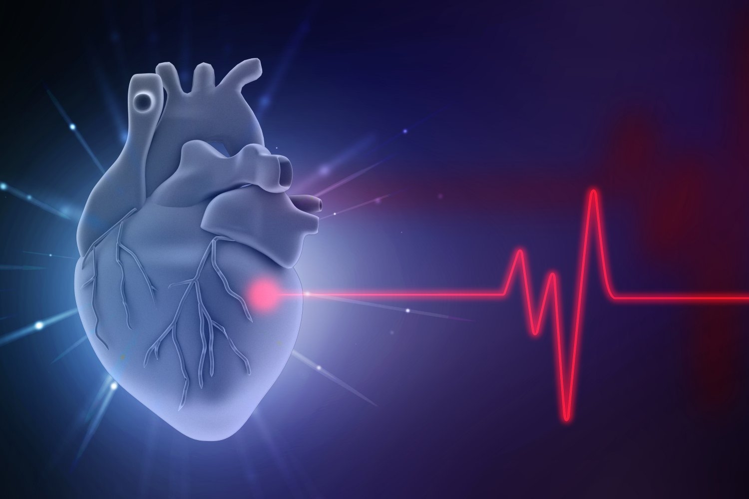
MIT and Harvard Medical School researchers have recently published free access paper in Nature communications medicineIntroducing a non -invasive in -depth learning approach which analyzes the signals of the electrocardiogram (ECG) to predict with precision the risk of a patient to develop heart failure. In a clinical trial, the model has shown results with precision comparable to standard but more invasive procedures, which gives hope to those who risk heart failure. The condition has recently seen A sharp increase In mortality, especially in young adults, probably due to the growing prevalence of obesity and diabetes.
“This document is a highlight of things that I have been talking about for other years for several years,” said the main author of the newspaper, Collin Sultz, director of Harvard-MIT program in health and technology sciences and affiliate of Mit Abdul Latif Jameel Clinic for automatic health learning (Jameel clinic). “The purpose of this work is to identify those who are starting to fall sick before you even have symptoms so that you can intervene early enough to prevent hospitalization.”
Of the four bedrooms of the heart, two are earrings and two are ventricles – the right side of the heart has an atrium and a ventricle, and vice versa. In a healthy human heart, these rooms operate in a rhythmic synchrony: the blood poor in oxygen flows into the heart via the right atrium. The right atrium contracts and the pressure generated pushes the blood in the right ventricle where the blood is then pumped into the lungs to be oxygenated. The blood rich in oxygen of the lungs then drains in the left atrium, which contracts, pumping the blood in the left ventricle. Another contraction follows and the blood is ejected from the left ventricle via the aorta, flowing into the veins branching towards the rest of the body.
“When the left auricular pressures become high, blood drainage of the lungs in the left atrium is hampered because it is a high pressure system,” says Stalltz. In addition to being a professor of electrical and computer engineering, Stultz is also an in -office cardiologist at Mass General Hospital (MGH). “The more the pressure in the left atrium is high, the more you develop pulmonary symptoms – shortness of breath and so on. Because the right side of the heart pumps the blood through the lung vascular system to the lungs, the high pressures of the left atrium result in high pressures in the pulmonary vasculure. “
The current gold stallion to measure left ear pressure is right cardiac catheterization (RHC), an invasive procedure which requires a thin tube (the catheter) attached to a pressure transmitter to be inserted into the right heart and the pulmonary arteries. Doctors often prefer to assess the risks in a non -invasive manner before resorting to the RHC, by examining the weight, the patient's blood pressure and the heart rate.
But from Stultz, these measures are gross, as evidenced by the fact that One in four heart failure is read in hospital within 30 days. “What we are looking for is something that gives you information like that of an invasive device, other than a simple weight scale,” explains Sultz.
In order to collect more complete information on a patient's heart disease, doctors generally use a 12 dishes ECG, in which 10 adhesive patches are stuck on the patient and linked to a machine that produces information from 12 different angles from the heart. However, ECG machines with 12 derivations are only accessible in clinical environments and they are not generally used to assess the risk of heart failure.
Instead, what Stultz and other researchers offer is a Hemodynamic cardiac monitoring system (CHAIS), a network of deep neurons capable of analyzing ECG data from a single track – in other words, the patient only needs to have a single adhesive, commercially available on their chest that they can bring outside the hospital, without an attack on a machine.
To compare the cellar with the current gold stallion, RHC, the researchers selected patients who were already planned for catheterization and asked them to wear the patch 24 to 48 hours before the procedure, although patients were invited to remove the patch before catheterization. “When you arrive less than an hour and a half (before the procedure), it's 0.875, so it's very, very good,” explains Sultz. “Thus, a measurement of the device is equivalent and gives you the same information as if you were catheted in the next hour.”
“Each cardiologist includes the value of left ear pressure measures to characterize heart function and optimize treatment strategies for patients with heart failure,” said Aaron Aguirre SM '03, PHD '08, cardiologist and intensive care doctor in MGH. “This work is important because it offers a non -invasive approach to estimate this essential clinical parameter using a widely available heart rate.”
AGUIRRE, who has finished a doctorate in medical and medical engineering in the MIT, expects that with additional clinical validation, the cellars will be useful in two key areas: first, this will help select patients who will benefit the most from a more invasive cardiac test via RHC; And secondly, technology could allow series monitoring and the monitoring of left atrial pressure in patients with heart disease. “A non -invasive and quantitative method can help optimize treatment strategies in patients at home or in hospital,” explains Aguirre. “I am delighted to see where the MIT team takes this.”
But the advantages are not limited to patients – for patients with heart failure difficult to manage, it becomes a challenge to prevent them from being reader in the hospital without permanent implant, taking more space and more time besieged medical workforce and under-effective.
Researchers have another current clinical trial using Chais with MGH and Boston Medical Center which they hope to conclude soon to start data analysis.
“In my opinion, the real promise of AI in health care is to provide state -of -the -art care for everyone, regardless of their socioeconomic status, their experience and their place of life,” explains Sultz. “This work is a step towards achieving this objective.”